
Any abnormal growth in the mouth and the surrounding tissue is considered an oral mass. The tissues involved can be maxillo-facial bones (maxilla, mandible, facial, nasal, zygomatic, temporal, palatal bones), the temporomandibular joint, or soft tissues like gums, tongue, cheeks, lips, tonsils, palate, salivary glands, oropharynx (the area in the back of the mouth), and regional lymph nodes.
What is An Oral Tumor?
In general, the term tumor applies to any type of abnormal growth. Tumors are categorized as benign or malignant based on their biological behavior.
A benign tumor is one that has a slower growth rate and does not metastasize to the regional lymph nodes or other organs. Even though a benign tumor is not necessarily fatal, it can have an aggressive growth pattern and can cause severe tissue damage and pain.
A malignant tumor (also called cancer or neoplasia) is a term reserved for tumors that grow fast and have the capacity to metastasize (spread to) lymph nodes and distant organs.
Growth-like lesions in the mouth can also be caused by non-neoplastic processes like inflammation, infection, foreign bodies, or trauma. These lesions can be confused with oral tumors and should be diagnosed as soon as possible after being noticed.
Conversely, oral tumors can present clinically as an ulcer or a non-healing extraction site, and not with a typical lump-like lesion.
There are numerous studies looking at the incidence rate of oral tumors, but in general, they represent about 6% of all tumors in dogs and 7.5% of all tumors in cats.
What Causes An Oral Tumor?
Just like in humans, oral cancer is caused by a combination of genetic and environmental factors–although the exact cause is unknown. Some cancers, however, are more frequently diagnosed in certain breeds, sex, or age-groups.
Periodontal disease, which is an inflammation of the, gums and supporting structures around the teeth, can cause non-neoplastic masses. It is the most common disease diagnosed in small animals. It may cause gingival growths in both cats and dogs, ulcerations on the oral mucosa, and loose teeth.
Trauma can also cause growths in and around the mouth. The absence of a tooth can cause an irritation followed by a growth in the soft tissues of the opposite arcade (pyogenic granuloma).
Immunologic disorders have been known to result in abnormal growths on the soft tissues of the mouth like eosinophilic granulomas. Another condition that is also multifactorial, stomatitis, can cause ulcers or growths in both cats and dogs.
Foreign bodies can become embedded in the tongue (burdock plant) or around the teeth, causing swelling. Infectious agents can cause a granuloma or an abscess, and allergic reactions can cause swellings of the mouth as well.
Other risk factors Include:
- Pigmented oral mucosa
- Exposure to excess sunlight in dogs
- Use of flea-collars
- Exposure to second-hand cigarette smoke
- Feeding canned food or tuna fish in cats
Types of Oral Tumors in Dogs & Cats
In dogs, malignant tumors were the most diagnosed pathology in biopsy samples submitted to the lab, found in about 37% of cases. Malignant melanoma, squamous cell carcinoma, and fibrosarcoma are the most common. Odontogenic tumors (arising from remnants of tooth-forming epithelium) are diagnosed in 34% of cases, and lesions of inflammatory origins in 28%.
In cats, lesions of inflammatory origin are the most diagnosed oral tumors—found in more than half of the biopsies (51-53%). The malignant oral neoplasms are diagnosed in 32-37% of cases, with squamous cell carcinoma being the most frequent, followed by fibrosarcoma, osteosarcoma and malignant melanoma.
Symptoms of Oral Tumors
The presence of a growth in the mouth can be easily overlooked until other signs of local discomfort become noticeable. A pet with an oral mass can have general signs of disease like lethargy, decreased appetite, weight loss, or signs specific to the oral cavity.
Most of the pets affected by oral tumors would have:
- Bad breath (halitosis)
- Excessive salivation
- Blood-tinged saliva
- Difficulty eating or selective appetite (will eat soft food but refuse kibble)
- Pawing at the mouth
- Dropping food from the mouth
- Abnormal tongue movements and pain on palpation
- When the throat is involved, there can be gagging, coughing, or breathing loudly
How Do We Diagnose Oral Tumors?
Not all oral masses are malignant, and not all malignant oral masses appear as growths. An accurate diagnosis is essential in determining the best course of treatment and the prognosis.
There are different diagnostic tests that should be performed together to gather all the necessary information. The investigation may start with a few blood tests, but everything else will require general anesthesia.
Diagnostic testing will start with a thorough oral examination and periodontal probing, dental X-rays, and computed tomography (conventional or cone beam computer tomography=CBCT). In rare cases, an MRI may be recommended.
Histopathological evaluation of a biopsy sample is of utmost importance. A tissue sample is collected from the tumor and sent to the pathology laboratory. The pathologist selected should have experience with oral tumors, as oftentimes they can be challenging.
Images of an oral tumor before surgery.
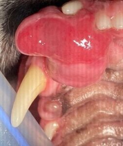
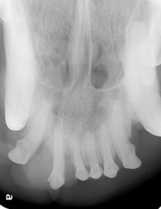
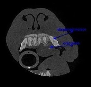
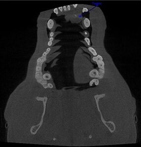
Oral tumors are staged based on clinical criteria adopted by the World Health Organization (WHO). The TNM system is assessing the size of the tumor (T), the lymph node involvement (N), and the presence or absence of metastasis (M). The addition of numbers to these letters indicates the extent or involvement of the neoplasm.
The term stage does not necessarily mean a regular progression from stage one to four, rather it is mostly used to define prognosis and treatment.
Early detection and diagnosis are key in reducing the morbidity and mortality associated with oral tumors. Preventive care through regular vet visits, yearly dental cleanings and thorough oral exams under anesthesia, and home dental care will all lead to an early diagnosis and allow prompt initiation of therapy.
Treating Oral Tumors
The treatment of choice for most oral tumors is surgical removal. The first procedure has the best chance of success, and it will be guided by the vital information collected during diagnostics.
Surgical removal with curative intent is performed by excising the mass with a portion of the healthy-looking tissue around it, called the margin. The recommended margins for excision will depend on the histopathological diagnosis.
A benign hyperplastic growth can benefit from marginal removal, a benign odontogenic tumor will require 1 cm margins, whereas for malignant tumors 2 cm margins are recommended.
The tumor is measured, and the margins are drawn with a sterile surgical marker. All tissues that fall within the marked line will be removed, including the bone and/or teeth.
In cases where the tumor is large and aggressive, extensive surgical procedures that remove a portion of the upper jaw (maxilla) or lower jaw (mandible) will need to be performed (maxillectomy or mandibulectomy). Surgeries performed with curative intent are usually more aggressive.
The tissue excised is then sent for histopathology to assess microscopic margins. The pathologist will measure the distance from the excised margin to the extent of neoplastic cells in the sample and inform the surgeons if the margins are complete or not. “Dirty” margins are a poor prognostic indicator.
Depending on the results, additional surgery or adjuvant therapy may be recommended.
Images of the same tumor after rostral maxillectomy, including the fragment excised.
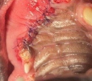
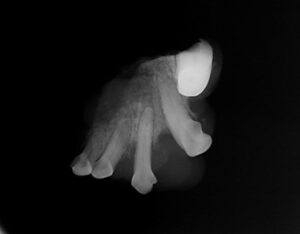
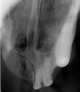
In some cases, the tumors are recognized and diagnosed late, and curative-intent surgery is no longer an option. These cases may sometimes benefit from palliative surgery, but the risk vs. benefit of a surgical procedure must be carefully considered.
Even extensive surgical procedures like a maxillectomy or mandibulectomy are well tolerated by dogs. Once they recover from anesthesia, they are able to go home, and most of them eat dinner the same night!
Cats are less tolerant of extensive surgeries, but with supportive care and assisted feeding, they can benefit from surgical treatment as well. A favorable outcome greatly depends on the staging of the tumor (type, size, lymph node involvement and metastasis at the time of diagnosis).
Non-Surgical Options
If surgery is not curative on its own, or the oral tumor is inoperable, adjuvant therapy like radiation or chemotherapy are an option. When the oral mass is non-neoplastic, the condition causing the growth may benefit from different treatment options.
Oral masses caused by periodontal disease will be treated with periodontal therapy, extractions of unsalvageable teeth, antibiotics, and anti-inflammatories.
In cases of feline stomatitis, partial or full-mouth extraction is the treatment of choice.
Other immune system disorders may require immunomodulatory medications (steroids, cyclosporine etc.). Trauma cases causing swelling are managed medically or surgically once the patient has been stabilized.
Board-Certified Vet in Phoenix
Early diagnosis of oral tumors in dogs and cats is critical to the health and wellness of your pet. That is why annual dental exams and cleanings are so important. If your pet is in need of a dental cleaning, contact us at Carefree Dentistry and Oral Surgery for Animals.
